Dr Helen Murray and Stanley Cardon
Our speakers at Tuesday’s were Dr Helen Murray and Stanley Cardon from the University of Auckland Centre for Brain Research.
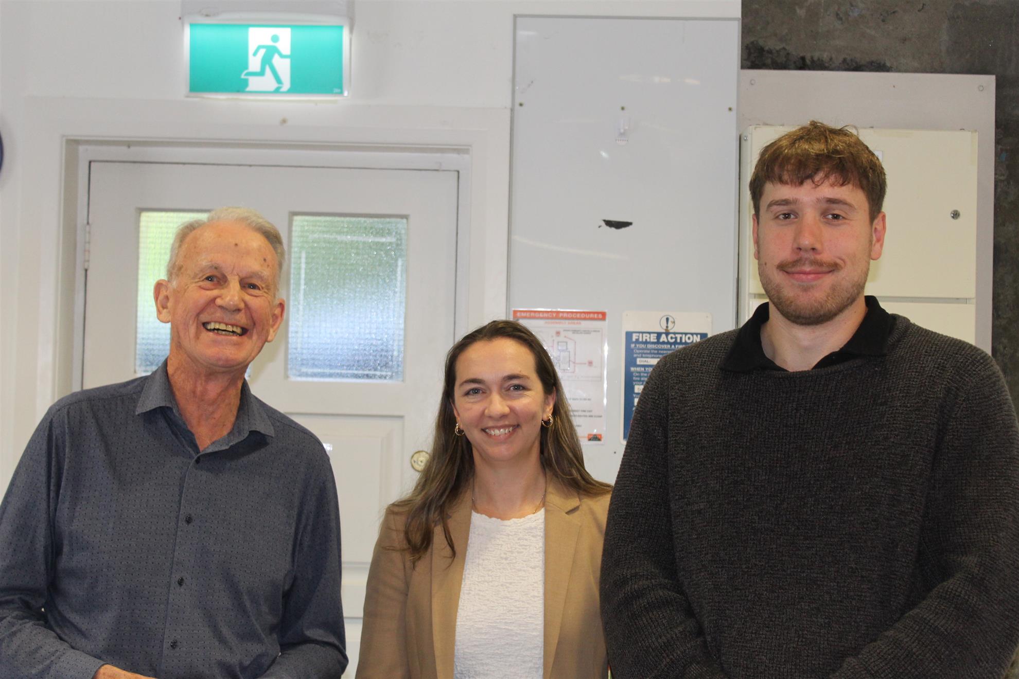
Their general theme was the impact of repeated head impacts and the link with dementia.
Helen opened the meeting by providing background on the research group's work and commenting on Stanley's master's highlights.
Helen explained the research on the link between repeated head impacts and the risk of developing dementia. Both she and Stanley have personal experience as they have engaged in collision sports, specifically ice hockey and rugby.
She introduced us to the concept of repeated head impact exposure, including concussions and other knocks during training or games. She noted that
athletes in contact sports can experience thousands of impacts annually, leading to significant exposure.
Helen talked about Health Risks and Clinical Syndromes. She noted the increased risk of cognitive decline and dementia in athletes exposed to repeated head impacts.
Dementia is described as cognitive change affecting daily life, caused by various brain diseases.
She mentioned specific conditions linked to repeated head impacts, such as chronic traumatic encephalopathy (CTE), Alzheimer's disease, and Parkinson's. The focus of the group that she leads and the centre for Braion research is on studying CTE due to its unique link to repeated head injuries.
Helen went on to talk about a recent study by New Zealand Rugby showing a 22% higher risk of neurodegenerative disease in former professional rugby players. The study also highlighted a 61% higher risk of Alzheimer's disease among rugby players compared to the general population. For some strange reason the risk is higher in backs than forwards, likely due to higher velocity collisions. It appears that longer playing careers increase the risk, indicating a significant issue in New Zealand.
Helen’s lab focus is on understanding how repeated head impacts affect the brain and developing methods to diagnose these conditions in living people. Biomarkers are crucial for identifying brain diseases like CTE, which are currently only diagnosable posthumously.
The goal is to monitor athletes over time, allowing for early intervention if signs of brain damage are detected. Biomarkers can be blood tests or MRIs, providing a measure of the likelihood of developing a brain disease.
Helen introduced us to biomarkers. The search for biomarkers begins with examining the brains of individuals with the disease being studied.
CTE is characterized by the accumulation of tau protein, causing neuron death and lesions at the bottom of the brain folds.
The condition progresses, with lesions moving down into areas involved in learning and memory. Blood vessels around the lesions show accumulation of tau protein, indicating damage from repeated head impacts.
Helen went on to talk about CTE affecting women and in non sporting contexts. She noted a case study from Australia shows the first case of CTE in a female, highlighting the non-sporting contexts of repeated head impacts. The study noted that female victims of domestic violence had CTE, demonstrating that the condition is not limited to athletes.
Helen noted the similarities between CTE and Alzheimer's disease, with both conditions causing brain cell loss. Multiple diseases can coexist in the same brain, complicating the study of biomarkers.
Helen told us that repeated head impacts cause the brain to move inside the skull, stretching and twisting brain folds. Areas at the bottom of the folds experience the most sheer stress and damage, leading to pathology. Her lab’s working hypothesis is that repeated injuries damage the blood-brain barrier, leading to inflammation and chronic changes.
The goal is to test this hypothesis and develop methods to identify conditions like CTE earlier.
Helen talked about the Blood-Brain Barrier and Inflammation. The blood-brain barrier is crucial for maintaining brain health by controlling what enters the brain. Repeated head impacts damage this barrier, leading to microhemorrhages and inflammation. Chronic exposure to these injuries can prevent the barrier from regenerating, leading to long-term damage.
The lab aims to study this cascade of damage to better understand and diagnose conditions like CTE.
Having laid the ground work Helen introduced Stanley Cardon who spoke to his
master's research related to the topic discussed above.
Stanley told us the lab uses multiplex immunohistochemistry to label dozens of proteins simultaneously on brain tissue.
Alzheimer’s samples show widespread astrocyte‑driven inflammation, while normal brains lack this pattern. CTE samples display focal inflammation that co‑localizes with tau‑rich lesions, unlike Alzheimer’s.
These distinct inflammatory profiles may help differentiate CTE from Alzheimer’s and suggest new therapeutic targets.
Researchers confirmed inflammatory markers in New Zealand CTE cases. The signature persisted in cases with co‑diagnosed Alzheimer’s disease.
Normal brains lacked the inflammatory pattern, validating its specificity.
Researchers identified NLRP1 as a reliable marker for CTE lesions.NLRP1 presence distinguishes CTE from other neurodegenerative diseases. MRI development aims to detect inflammation in living patients.
Helen concluded the address by telling us researchers are using rams to replicate brian injury in humans. Rams head but for social dominance in a flock. The effect of head buts is similar to repeated head knocks in humans.
This because sheep have a folded brain structure similar to humans, making them suitable for recapitulating CTE pathology.
The lab is collaborating with Massey University and the University of Auckland to validate the sheep model.
The study aims to understand the time course of CTE development and the impact of head injuries on sheep brains.
The pilot study involves monitoring sheep behavior and brain changes using MRI and histology. The goal is to quantify the impact of head butting on sheep brains and correlate it with pathological changes.
The research team includes experts in sheep behavior, MRI, and histology, ensuring comprehensive analysis. The long-term vision includes using the sheep model to test treatments and develop better diagnostic tools for CTE. Researchers will attach accelerometers, GPS, and video to sheep to quantify head‑butting impacts.
MRI and blood analyses will assess early brain changes and potential biomarkers.
It will be interesting to see the outcome of the pilot study.
Helen and Stanley were thanked by Alan Hayward.


